

Microscopy Gallery
During my PhD, I captured images that reveal beauty hidden at the cellular level.
This gallery provides a glimpse.
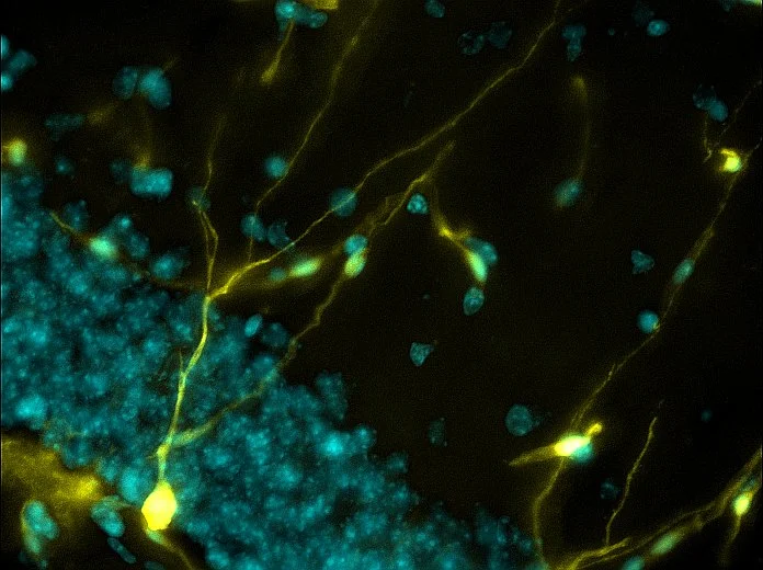
Genetically labeled neurons (yellow) represent neurogenesis (the birth of new neurons) and illustrate the brain's capacity for growth.
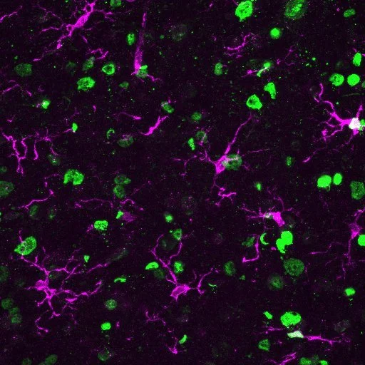
After a spinal cord injury, innate immune cells (magenta) overlap with fat molecules (green), suggesting that this molecule is involved in regulating inflammation.
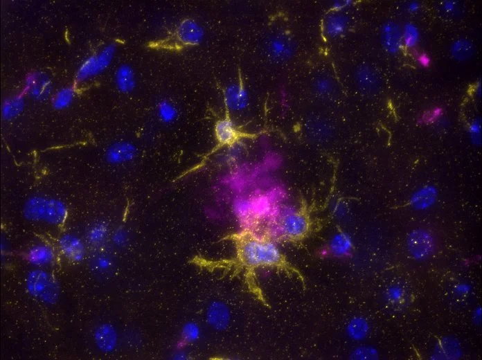
Immune cells in the brain, microglia (yellow), "clean up" plaques (magenta) associated with Alzheimer's Disease. The brain's immune cells are activated in neurodegeneration.
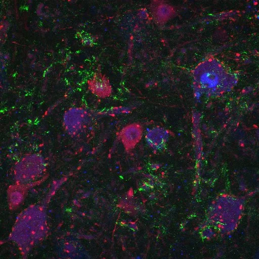
Complement protein C1q (green) aggregates on degenerating motor neurons (magenta) after spinal cord injury.
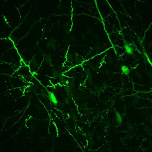
Mouse cortical neurons (green) were labeled with a virus, providing insights into brain and spinal cord motor control mechanisms.
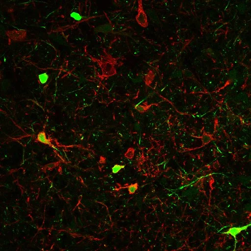
Motor control neural pathways that originate from the brain (green) and project to the spinal cord (red) overlap in the brain stem neurons (green & red).
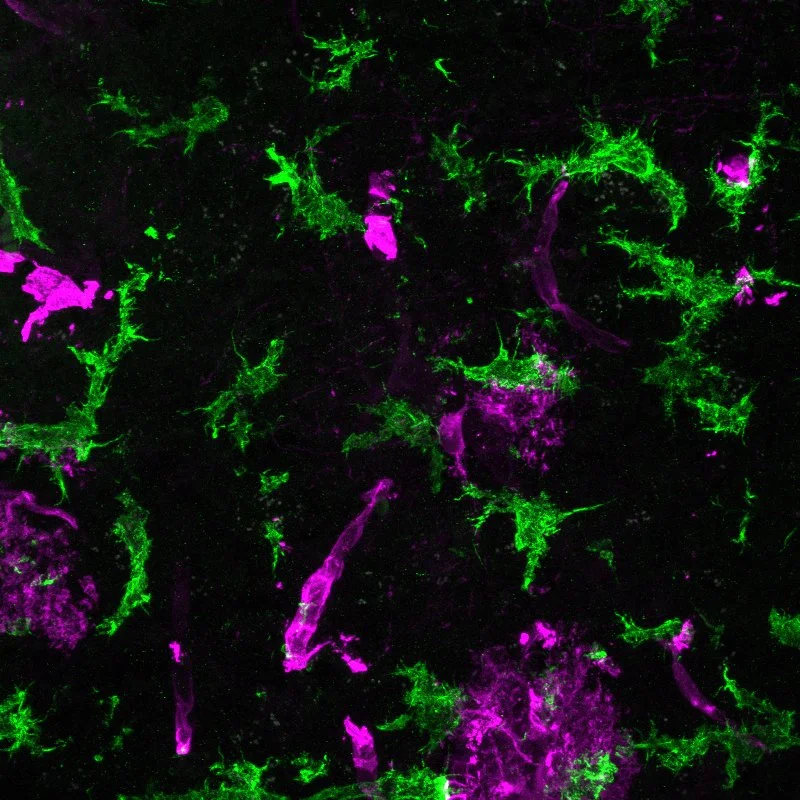
After a stroke, immune cells, microglia (green), become reactive.
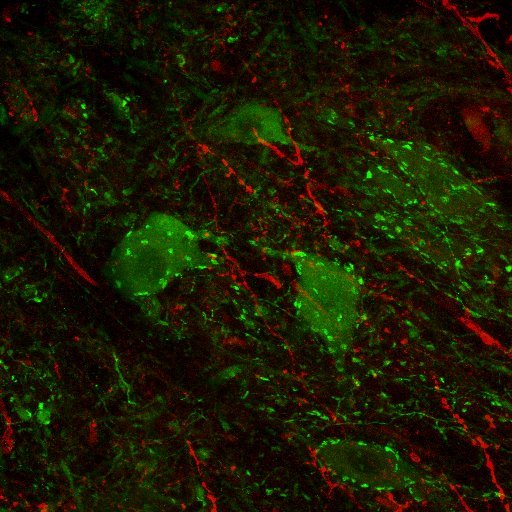
In the spinal cord, tiny excitatory connections, glutamatergic synapses (red), communicate with motor neurons (green).

Neurons virally traced from the brain stem (magenta) and pathways traced from the spinal cord (cyan) highlight a neuron population dedicated to forelimb function.

Motor neurons (pink), immune proteins (cyan), and neurons (yellow) in a whole spinal cord section after an injury.