

Microscopy Gallery
During my PhD, I captured images that reveal beauty hidden at the cellular level.
This gallery provides a glimpse.
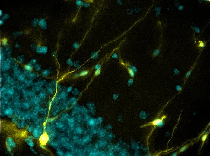
The Brain's Boundless Growth: Genetically labeled neurons (yellow) signify neurogenesis (the birth of new neurons) and illustrate the brain's capacity for growth.
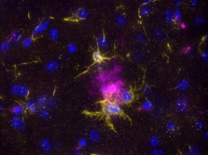
A Symphony of Immune Responses: After a spinal cord injury, innate immune cells (magenta) overlap with fat molecules (green). This suggests an active role of this fat molecule in regulating inflammation.
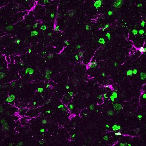
Plaquing Brain: Immune cells in the brain, microglia (yellow), "clean up" Alzheimer's Disease associated plaques (magenta). This illustrates the brain's defense mechanisms and highlights microglia as a potential target for therapeutic intervention.

Complementing Degeneration: Complement protein C1q (green) aggregates on degenerating motor neurons (magenta) after spinal cord injury.
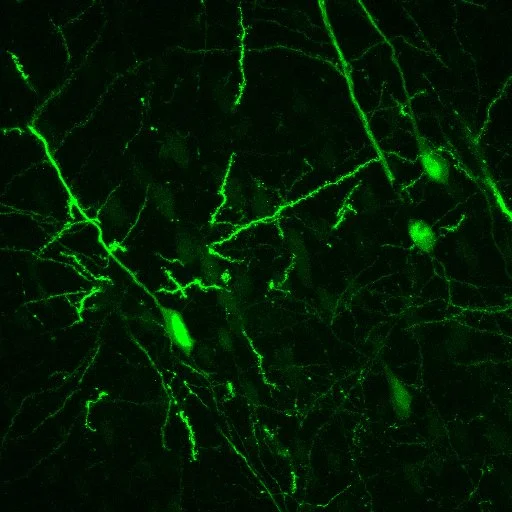
Tracing Movements: Mouse cortical neurons (green) were labeled with a virus, providing insights into neural circuitry and motor control mechanisms.

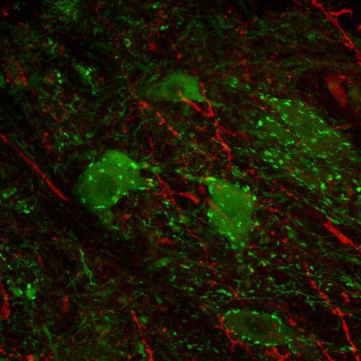
Where Pathways Merge: The convergence of neural pathways from the brain to the spinal cord (red) and from the spinal cord to the brain (green) traces the group of brain stem neurons (green & red) involved in forelimb motor control.

Microglia in Motion: In stroke, immune cells, microglia (green), are reactive, as indicated by genetically labeled blood vessels and astrocytes (magenta) expressing a stem cell marker.
Synaptic Conversations: In the spinal cord, tiny excitatory connections, glutamatergic synapses (red), communicate with motor neurons (green).

Forelimb Maestros: Neurons virally traced from the brain stem (magenta) and pathways traced from the spinal cord (cyan) highlight a neuron population dedicated to forelimb function.

Making the Right Moves: Motor neurons (pink), immune proteins (cyan), and neurons (yellow) in a whole spinal cord section after an injury.